
Reading Between the Genes: A Full-Length View of Glioblastoma at Single-Cell Resolution
A cancer that has resisted a generation of progress
Glioblastoma (GBM) remains the most aggressive primary brain tumour in adults, and despite decades of research and a standardised treatment approach comprising surgical resection, radiotherapy, and chemotherapy with temozolomide, the median survival is only about 15 months. The poor prognosis has changed little over a generation, leaving patients and their families devastated by the diagnosis.
Behind every one of those statistics is a person, and the patient must remain at the centre of everything we do. In our clinics, we meet families at what is often the most frightening moment of their lives, and we are asked the same question in different words: what can you do for me? For too long, the honest answer has fallen short of what we, as clinicians and scientists, would wish it to be. Surgery can debulk the tumour, radiotherapy and temozolomide can slow it down, but glioblastoma almost always returns, and when it does our options narrow sharply. Incremental gains are no longer enough; the boundaries of what is currently possible must be pushed.
Two features of GBM make it uniquely difficult to treat, and understanding them helps explain why progress has been so challenging. First, no two tumours look alike; even within a single tumour, cells adopt strikingly different molecular identities, a phenomenon known as intratumoural heterogeneity. Second, GBM cells extensively rewire how their genes are read, producing distorted messenger RNA (mRNA) molecules that give rise to abnormal or oncogenic proteins. This process, called alternative splicing, is emerging as one of the most consequential yet least understood drivers of the disease, and it has largely escaped detection by the technologies that dominate mainstream cancer genomics.
Our recent study, published in Nature Communications, was designed to confront both problems at once. By combining long-read sequencing (which captures full-length mRNA molecules) with single-cell resolution (which examines each cell individually rather than averaging signals across thousands), we set out to build the most comprehensive map to date of the "isoform landscape" of glioblastoma. The goal was straightforward: to identify the molecular features that truly distinguish a patient's tumour cells from their healthy cells, and in doing so to uncover treatment opportunities that conventional approaches have missed. That level of resolution is, in our view, what patients now require of us.
Why isoforms matter
Every human gene can, in principle, be spliced into multiple different mRNA molecules, each encoding a slightly different protein. These variants are called isoforms. In healthy cells, splicing is finely regulated. In cancer cells, splicing descends into chaos, producing isoforms that promote tumour growth, evade the immune system, or resist treatment.
Until recently, cancer transcriptomics has largely relied on short-read sequencing, which only reads fragments of each mRNA. To resolve full-length isoforms, one needs long-read sequencing using technologies that can read entire mRNA molecules from end to end. Applying long-read sequencing at the level of individual cells is a considerable technical challenge, but doing so provides something extraordinary: the ability to see, for each cell within a tumour, exactly which version of each gene is being expressed.
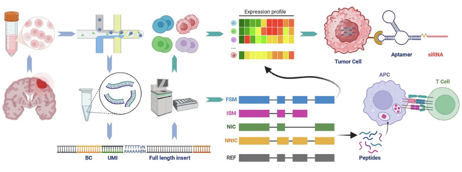
Figure 1. An overview of the study workflow. Tumour tissue from glioblastoma patients was dissociated into single cells, from which full-length RNA molecules were captured and read using two complementary sequencing technologies, short-read sequencing to establish cell identity, and long-read sequencing to resolve the entire structure of each RNA transcript. This combined approach allowed us to identify tumour-specific RNA isoforms and to nominate two classes of therapeutic opportunity: aptamer-based agents that engage both surface and intracellular targets on tumour cells, and peptide neoantigens that could be developed into personalised cancer vaccines.
Building a single-cell, long-read atlas of GBM
Using 27 surgical GBM specimens from patients treated at Queen Mary Hospital, Hong Kong, we generated matched short- and long-read single-cell RNA sequencing data from more than 182,000 cells. This produced, to our knowledge, the largest isoform-resolved atlas of human GBM assembled so far.
Three broad findings emerged.
-
Widespread isoform rewiring in tumour cells. Nearly 100,000 mRNA transcripts were differentially expressed between tumour and non-tumour cells. Remarkably, over 6,600 genes produced protein-coding isoforms exclusively in tumour cells, while their non-cancerous counterparts produced non-functional versions. Many of these switches occurred without any change in overall gene expression, meaning they would have been entirely invisible to conventional analyses. A striking example is NTRK2, which encodes the neurotrophin receptor TrkB. In one particularly aggressive tumour subtype, the mesenchymal-like state, cells preferentially expressed a truncated form of TrkB lacking the intracellular signalling domain. This truncated isoform, known from previous work to enhance glioma growth, is essentially invisible at the gene level but jumps out clearly at the isoform level.
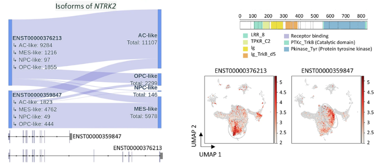
Figure 2. A single gene, NTRK2, produces two very different RNA isoforms in glioblastoma. The full-length isoform (ENST00000376213) predominates in astrocyte-like tumour cells, while a shorter isoform (ENST00000359847) that lacks the intracellular signalling domain predominates in the more aggressive mesenchymal-like tumour cells. The two maps on the right show where each isoform is expressed across thousands of individual cells from the tumour, with red indicating high expression. This example illustrates a broader principle of the study: biologically important differences between tumour cell populations are often invisible at the gene level and only become apparent when RNA is read at full length.
- A framework for dual-targeting precision therapies. One of the great frustrations in GBM treatment is that most tumour-associated proteins are also expressed, at some level, in healthy tissue, making it difficult to develop treatments that spare normal cells. We reasoned that tumour-specific isoforms might offer a way around this: even when the underlying gene is expressed in healthy cells, the particular isoform seen in tumour cells may be unique. Building on this idea, we created an analytical framework to identify pairs of tumour-specific isoforms that are present only in cancer cells, both inside and outside the cells. Surface targets could be recognised by aptamers, small nucleic acid molecules that cross the blood-brain barrier more effectively than antibodies. This opens the possibility of using aptamer-drug conjugates as therapy, a new type of treatment that can target both cancer cells specifically and the brain, making it particularly promising for brain cancer. Applying this framework, we identified 40 highly patient-specific target pairs across our cohort. Each pair was virtually absent from the ~48,000 healthy tissue samples in the GTEx reference atlas.
-
Thousands of "hidden" isoforms, and a new source of cancer vaccine targets. Because long-read sequencing reads mRNAs end-to-end, it can reveal transcripts that are entirely absent from existing reference annotations. We identified 6,524 such high-confidence novel isoforms in GBM, 179 of which were tumour-specific.
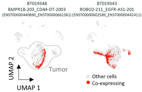
Figure 3. Each glioblastoma patient carries a distinct combination of tumour-specific RNA isoforms that could be exploited therapeutically. Shown here are two representative patients, each with a different pair of targets, one on the tumour cell surface and one inside the cell, that are co-expressed almost exclusively by their tumour cells (highlighted in red) and are absent from healthy cells. Pairs of this kind are candidates for a dual-specific therapeutic strategy in which a surface-binding aptamer delivers a gene-silencing agent to intracellular targets, allowing precise action against the tumour while sparing healthy tissue. The distinctiveness of each patient's target pair underscores the need for personalised design in future glioblastoma therapies.
We then asked, could these tumour-exclusive isoforms give rise to small peptides that the immune system might recognise as "foreign"? Using two independent prediction algorithms and validating with a third, we identified 89 isoforms encoding peptides with strong predicted binding to human leukocyte antigen (HLA) class I molecules, the molecular platforms our immune system uses to present abnormal proteins to T cells. Importantly, several of these predicted neoantigens showed broader and stronger predicted immune presentation than well-known mutation-derived neoantigens that have already entered peptide-vaccine clinical trials. This is particularly meaningful for GBM, a cancer with a notoriously low mutational burden, which has historically limited the effectiveness of immunotherapy. Splicing-derived neoantigens may therefore represent an untapped and abundant source of tumour-specific targets, a rich seam of "foreignness" that a personalised vaccine could exploit.
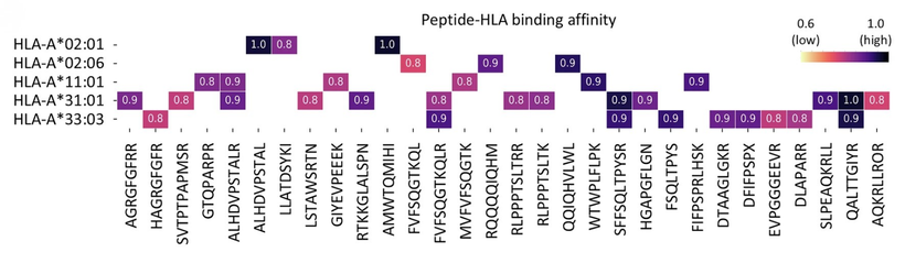
Figure 4. Candidate neoantigens, short protein fragments derived from tumour-specific RNA isoforms, ranked by their predicted ability to be presented to the immune system through the five HLA class I molecules most common in East Asian populations. Darker colours indicate stronger predicted binding, and therefore a higher likelihood that the fragment could be recognised by T cells and used to trigger an anti-tumour immune response. These peptides are not present in the normal human proteome, making them attractive targets for the design of personalised cancer vaccines against glioblastoma.
From bedside to bench: a collaboration framework
Work of this kind depends on infrastructure that no single laboratory can assemble on its own, and the study drew on two partnerships that have matured in Hong Kong in recent years.
The tumour specimens were contributed through the Clinical Neuroscience Consortium (CNC), established by the LKS Faculty of Medicine, The University of Hong Kong (HKUMed) in collaboration with Queen Mary Hospital. The CNC operates under a shared governance and biobanking framework that links neurosurgeons, neuro-oncologists, neuropathologists and translational scientists, allowing tissue to be collected, annotated and released for research with the clinical detail required for isoform-level analysis. The Brain Tumour chapter of the CNC, working with the Division of Neurosurgery at HKUMed and Queen Mary Hospital, coordinated the intraoperative sampling and clinical annotation on which this study depended.
The sequencing and computational analysis were carried out in partnership with the Hong Kong Genome Institute (HKGI), the operational arm of the Hong Kong Genome Project. HKGI provided the long-read platforms and the informatics environment needed to perform single-cell isoform analysis at this scale, a combination that is not yet routinely available in most academic settings. Our collaboration with HKGI extends beyond this project into a broader programme of genomic and transcriptomic characterisation across several malignancies, with the shared aim of translating population-scale sequencing capacity into direct patient benefit. The alignment of clinical services, academic research, and public-sector genomic infrastructure that made this study possible remains uncommon internationally, and continued investment in this alignment will determine how quickly work of this kind can move from single studies into standard practice.
What comes next
The results of this study lay a foundation, not a conclusion. Three questions now stand between the biology we have described and the patients we hope will benefit from it. First, do the predicted neoantigens actually elicit T-cell responses in patients? Answering this will require systematic immunological validation, ideally embedded within early-phase clinical studies. Second, can aptamer-drug conjugates directed at the patient-specific isoform pairs we identified achieve meaningful efficacy in preclinical models, and with an acceptable therapeutic window? Third, and perhaps most demanding, how can a fundamentally personalised approach, in which each patient carries their own target pairs and their own neoantigens, be delivered through a workflow that hospitals and health systems can realistically operate?
Work on all three fronts is already underway in our laboratory and with our immediate collaborators. Immunogenicity studies of the predicted neoantigens, preclinical development of aptamer-drug conjugates against the isoform pairs we identified, and early design work on a clinically deployable personalised workflow are each being taken forward in parallel, so that the biological, chemical and operational arms of an isoform-guided strategy can mature together rather than sequentially. We are now extending these efforts beyond our immediate setting, building the regional and international collaborations that a personalised approach of this kind will ultimately require.
These are substantial questions, but they are not without precedent. The rapid clinical progress of mRNA-based personalised cancer vaccines in melanoma and pancreatic cancer has shown that patient-specific target identification, manufacture and administration can be compressed into clinically viable timeframes. Parallel advances in aptamer chemistries in other disease settings suggest that the delivery arm of an isoform-guided strategy is similarly within reach. What has been missing, until now, is a molecular map at the resolution needed to define the targets themselves. That is what this study begins to provide.
For a disease as aggressive as glioblastoma, incremental gains are no longer enough. Reading the transcriptome in its full-length, single-cell complexity has revealed a class of vulnerability that short-read approaches were never positioned to see. Turning that vulnerability into benefit for patients is the work that now lies ahead, and it is work that no single group will complete alone. We look forward to advancing it together with colleagues across the region and internationally, and to doing so from HKUMed, Queen Mary Hospital, and the Hong Kong Genome Institute, supported by the University Grants Council and the Research Grants Council, which is well configured to move discoveries of this kind toward the clinic.
Original Publication
Tang, W., Lo, C.W.S. et al. Mapping glioblastoma's isoform diversity using long-read single-cell analysis. Nature Communications 17:5640 (2026). https://doi.org/10.1038/s41467-026-72258-2
Authors
Prof Aya El Helali, Clinical Assistant Professor, Department of Clinical Oncology and Centre of Cancer Medicine, School of Clinical Medicine, Li Ka Shing Faculty of Medicine, The University of Hong Kong; Steering Committee Member, Clinical Neuroscience Consortium, Li Ka Shing Faculty of Medicine-Queen Mary Hospital
Prof Brian HY Chung, Interim Chief Executive Officer and Chief Medical and Scientific Officer, Hong Kong Genome Institute; Clinical Associate Professor, Department of Paediatrics & Adolescent Medicine, School of Clinical Medicine, Li Ka Shing Faculty of Medicine, The University of Hong Kong
Professor Gilberto Leung, Clinical Professor, Department of Surgery, School of Clinical Medicine, Li Ka Shing Faculty of Medicine, The University of Hong Kong, Convenor, Clinical Neuroscience Consortium, Li Ka Shing Faculty of Medicine-Queen Mary Hospital
All figures reproduced from the original publication under CC BY-NC-ND 4.0.